DENTAL DIAGNOSTICS
Modern technology guarantees the best possible diagnostics. It enables your dentist to come up with the ideal treatment plan.
DIGITAL INTRAORAL RADIOGRAPHY AND RVG RADIOVISIOGRAPHY
Digital intraoral x-ray systems have a very short exposition, which means that they produce minimum radiation. RVG radiovisiography is used to transfer x-rays onto a computer screen.
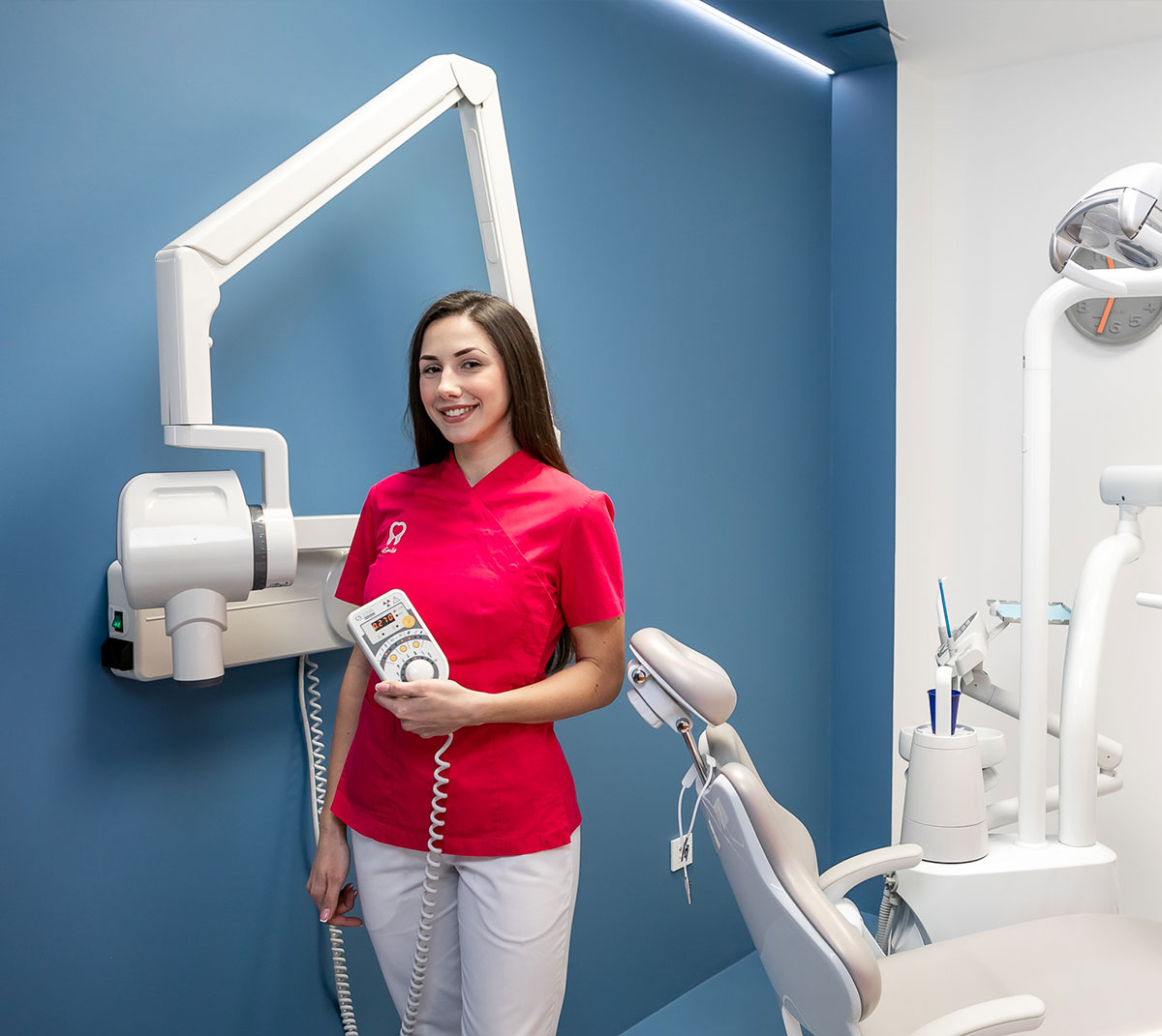
4Smile Dental Center uses the latest Carestream digital intraoral camera – a low-frequency x-ray system with an extremely short exposition that produces minimum radiation, along with an RVG sensor for the digital transmission of such images onto a computer screen.
The RVG is a digital sensor which enables us to make an intraoral x-ray without the use of film.
This sensor completely replaces conventional dental film. It allows us to make periapical, occlusal and bitewing x-rays and immediately display them on a computer screen. Setting the right diagnosis is faster and more efficient using a digital camera, which has a much higher resolution than the classic, film based radiograph.
Radiation from a digital intraoral camera is 90% lower compared to standard film.
Taking a clear x-ray only takes a fraction of a second, which is much less harmful for the patient. If it is necessary, it is possible to take multiple images at once.

DIAGNOSIS IS THE FIRST STEP TOWARDS A HEALTHY SMILE
PAN – PANORAMIC RADIOGRAPH (ORTHOPANTOMOGRAM)
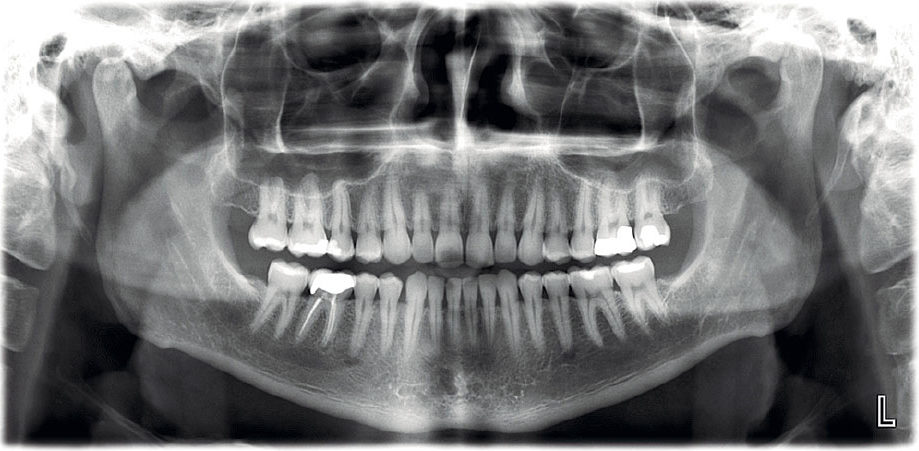
An orthopantomogram is a panoramic dental x-ray of both jaws. It is the principle diagnostic tool in dentistry and a crucial factor in treatment planning.
An orthopantomogram provides an x-ray image of both jaws.
Alongside clinical examination, a panoramic digital x-ray is the basic diagnostic tool in today’s dentistry.
Your panoramic x-ray should not be older than 6 months.
CBCT – A 3D IMAGE OF THE JAW (CONE BEAM COMPUTED TOMOGRAPHY)
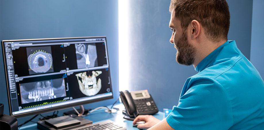
A CT of the jaw is a 3D x-ray image vital for planning any surgical procedure, especially implant placement.
A CBCT of the jaw provides a highly detailed, three-dimensional image of the jaw which allows more precise planning of a surgical (implant) procedure. 3D imaging is also increasingly used for establishing endodontic diagnoses.


